Individual Laboratories
The following facilities are available to researchers across the MSU campus as well as outside of the MSU community.
During 2002-2003, classrooms 2125 and 2135 (approximately 4,000 square feet) were renovated and converted to very high quality biological wet laboratory space. The adjacent classroom 2150 has been identified for renovation to laboratory space to complete the new facility. The cost for renovation was about $1.5 million. This new facility consists of wet lab space for tissue scaffold fabrication and characterization, a separate cell culture room for culture, testing and maintenance of cell lines that currently include osteoblasts, hepatocytes, neuronal cells and fibroblasts, and a separate Class 10,000 clean room, which will house equipment necessary for biomedical engineering device fabrication.
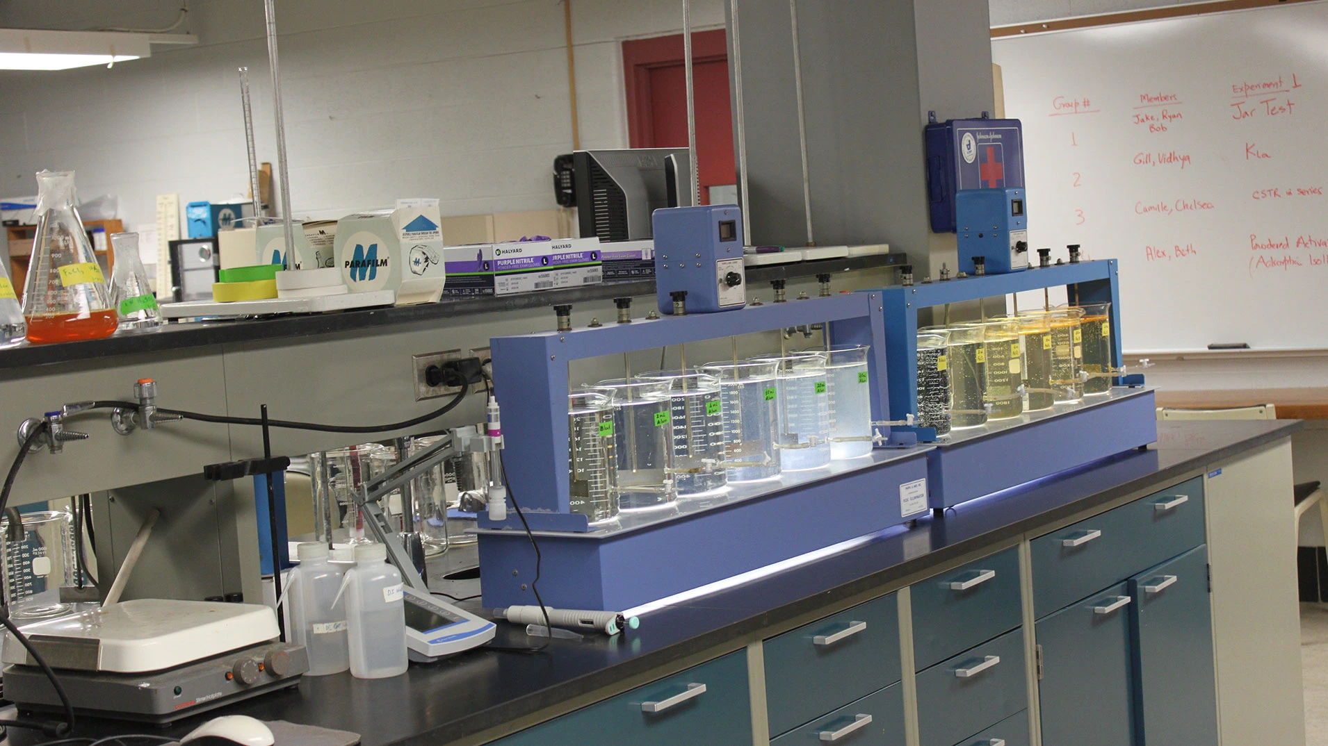
Clean, well-controlled, sterile conditions are requisite for cell and tissue culture experiments. Six separate hoods are provided in the tissue culture room for specific cultures including osteoblasts, hepatocytes, fibroblasts and neuronal cells. The Laboratory will also house equipment necessary to support the characterization of cellular function such as an HPLC, an electrokinetic sonic amplitude analyzer, plate readers, electrophoresis equipment, spectrophotometer, incubators, autoclave, centrifuges, optical and fluorescence microscopes, nitrogen storage tank for cell lines, -80ºC freezer, -20ºC freezer and refrigerators. This equipment is specialized for cell work and great care will be taken to prevent contamination. Often experiments run for several weeks and it is vital that the lab environment be controlled and monitored in order to collect long-term data. In part to protect our ability to monitor this lab, security devices were installed that will enable us to allow only necessary personnel into the lab.
Engineering faculty in collaboration with faculty from the departments of Physiology, Microbiology, Biochemistry, Chemistry and Small Animal Clinical Science/Orthopedics will use these facilities for their research activities.
Contact Kris Chan, krischan@egr.msu.edu, (517) 432-4530.
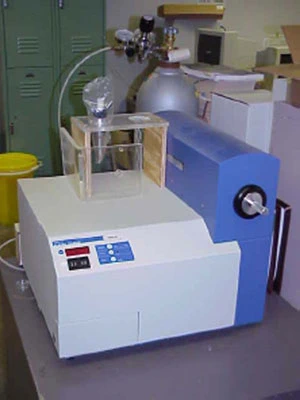
Within the Department of Chemical Engineering and Materials Science, there are 6 creep frames and one state-of-the-art thermomechanical testing machine capable of performing experiments at temperatures up to 1200 C and loads up to 10kN in vacuum (10-8 torr), inert gas, or air environments.
Contact Carl Boehlert, boehlert@egr.msu.edu, (517) 353-3703.
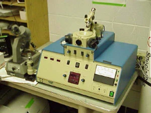
The Composite Materials and Structures Center is one of the largest integrated facilities for polymer and composites research and development in a non-industrial environment. With $9 million in equipment, the facility provides processing equipment and rheological, mechanical, adhesion, and surface evaluation.
Contact Lawrence Drzal, drzal@egr.msu.edu, (517) 353-7759.
Electron Microscopy Facilities include a scanning electron microscope and a 200kV transmission electron microscope. Microanalytical characterization is available using energy dispersive x-ray analysis while micro crystallographic analysis can be carried out using electron back scattering diffraction and/or selected area electron channeling patterns. The department also has equipment for X-Ray diffraction. The facilities are available to researchers from across campus and from outside the MSU community.
Contact Prof. Martin Crimp at crimp@egr.msu.edu or (517) 355-0294.
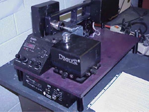
A Protein Expression Laboratory was funded by the Michigan Life Sciences Corridor. The mission is to use genetically engineered microbes to manufacture proteins needed for research. The lab has two 10-L computer controlled bioreactors and plans to add a third, 100-L bioreactor in 2003.
Contact Mark Worden, worden@egr.msu.edu, (517) 353-9015.
Electron Microscopy and X-Ray Diffraction Facilities
Electron Microscopy Facilities include a scanning electron microscope and a 200kV transmission electron microscope. Microanalytical characterization is available using energy dispersive x-ray analysis while microcrystallographic analysis can be carried out using electron back scattering diffraction and/or selected area electron channeling patterns. The department also has equipment for X-Ray diffraction. The facilities are available to researchers from across campus and from outside the MSU community.
For access to the facilities and/or additional information, please contact Prof. Martin Crimp at crimp@egr.msu.edu or (517) 355-0294.
Electron microscopes
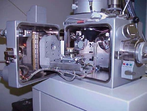
CamScan 44FE Field Emission Gun Scanning Electron Microscope.
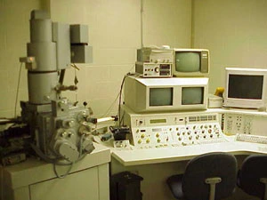
CamScan 44FE Field Emission Gun Scanning Electron Microscope
Uniquely configured to carry out a wide range of materials studies including crystallographic analysis using ECCI, SACP, and EBSD.
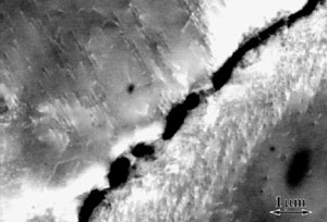
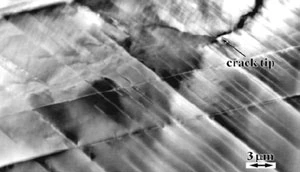
ECCI image of dislocations along a crack edge in a 4-point bend specimen of NiAl.
Instrument features
Secondary electron resolution of 2.5 nm at 25 kV and 5.0 nm at 5 kV.
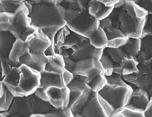
Secondary electron image of a fracture surface of Fe-50at% Al.
Backscattered electron resolution of 5.0 nm at 25 k.
Backscattered electron image showing channeling contrast from twins in TiAl.
Oversize chamber with fully motorized stage for automated data collection. Specimens as large as 3 kg can be observed with a stage x-and-y travel of 100 mm x 100 mm. In-situ mechanical testing using tensile, bending, and fatigue stages.
The oversize chamber accommodates large specimens and a variety of in-situ stages.
Selected area channeling pattern capabilities (SACP) with a spatial resolution of 3 micrometers.
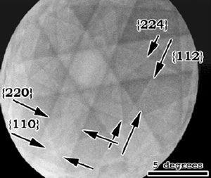
Selected area channeling pattern from TiAl reveals superlattice line details.
Electron back scattered diffraction/orientation imaging microscopy (EBSD/OIM) capabilities using a CamScan Ortex camera and HKL Technologies Channel Software. System allows both beam and stage scanning control.
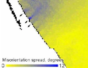
EBSD orientation map shows crystal rotation and dislocation slip band at grain boundary in gamma-TiAl.
The characterization facilities are supported by a wide range of state-of-the-art sample preparation facilities suitable for researchers working with metals, ceramics, polymers, and composites.
Major equipment
Fishione model 1020 plasma cleaner - Both TEM and bulk SEM sample holders are available.
Gatan precision ion polishing system (PIPS) - Low angle milling system with variable sector control.
VCR group 500i electronically dampened dimpler - Precision dimpler for both TEM thin foil and bulk controlled thinning.
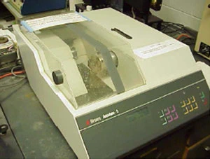
Struers ACUTOM 5 microprocessor controlled high speed wafering saw - Multiple controlled thin sections can be automatically sliced.
Other support facilities
- Struers Tenulpol - 3 dual jet electropolisher
- South Bay Technologies single sided jet electropolisher
- South Bay Technologies 515 dimpler
- South Bay Technologies 350 slurry dick cutter
- Gatan 601 ultrasonic disk cutter
- Denton vacuum DV-502 bell jar evaporator
User training for our various analytical instrument takes place through a number of MSU courses offered through the department, including:
- MSE 481 Spectroscopic and Diffraction Analysis of Materials
- MSE 870 Electron Microscopy in Materials Science
- MSE 881 Advanced Spectroscopy and Diffraction Analysis of Materials
In addition to formalized course training, individual training can be arranged with our technical staff. This is particularly useful for researcher needing assistance with either scanning electron microscopy or sample preparation.
CamScan 44FE
MSU users: $18.00/hr
Other non-profits: $45.00/hr
For-profit entities: $130.00/hr